Anatomy
This is as much as you need to know about vocal anatomy, but it's simplified. If you want more detail, check out an anatomy textbook.
Cartilages and Muscles of the Larynx
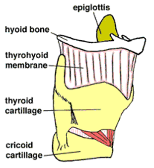
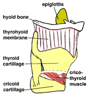
Figure 4: In the picture on the right, part of the thyroid cartilage is cut away to show the extent of the cricothyroid muscle, inserting into the inside of the thyroid cartilage.
Anatomy of the Larynx
- Cricoid (rhymes with "thyroid") cartilage - As the top ring of the trachea, the cricoid cartilage is shaped like a signet ring, wider in the back than the front.
- Thyroid cartilage - the thyroid cartilage fits over the cricoid cartilage, and is hinged so that it can slightly rock forward and downward. The thyroid cartilage comes to a point in the front; this point is termed the thyroid notch, but is commonly called the Adam's Apple. The vocal folds attach at the inside of the thyroid notch.
- Arytenoid (pronounced ah-RIHT-uh-noid) cartilages - These sit atop the back of the cricoid cartilage and hold the back end of the vocal folds. The arytenoid cartilages can rock, glide, and pivot, thus controlling the movement of the vocal folds.
- Vocal Folds (Vocal Cords) - These remarkable structures provide a valve for the airway and also vibrate to produce the voice. The vocal folds are multilayered structures, consisting of a muscle covered by a mucosal covering.
- Glottis - This is the space between the two vocal folds. When the vocal folds adduct, the glottis closes; when the vocal folds abduct, the glottis opens. The adjectives "glottal" and "glottic" are used to describe many aspects of vocal fold movement. The glottis opens and closes during vibration. Refer to the corresponding pictures.
- Epiglottis - This soft cartilage serves as part of the protective swallowing mechanism. It folds backward over the glottis during a swallow so that food and liquids do not go into the lungs. It is not involved in normal voice production.
- Hyoid (rhymes with "thyroid") Bone - This horseshoe-shaped bone is positioned slightly above the thyroid cartilage and is the only bone in the body not connected to any other bone. The hyoid bone provides the attachment for many of the muscles of the tongue, jaw, and neck.
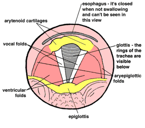
Figure 5: This is a view from above the larynx. In this view, mucosa covers the muscles and cartilage.
Muscles
Intrinsic Laryngeal Muscles
These are the muscles within the larynx itself.
All these muscles are paired. That is, they are symmetrically arranged on the left and right sides of the larynx.
- Posterior cricoarytenoid - These are the only muscles involved in abduction. They open the glottis by pulling the back ends of the arytenoid cartilages together. This pulls the front ends (where the vocal folds attach) apart, therefore pulling the vocal folds apart.
- Lateral cricoarytenoid - These are adductors. They close the glottis by pulling the back end of the arytenoid cartilages apart. This pulls the front ends together, making the vocal folds come together.
- Thyroarytenoid - These are the muscles that form the body of the vocal folds themselves. They shorten the vocal folds by pulling the arytenoid (back) end of the vocal folds toward the thyroid (front) end. This shortens the vocal folds and bunches them up, which causes them to vibrate more slowly, thus lowering pitch. The thyroarytenoid muscles also have a force to strengthen glottic closure. That is, they help bring the vocal folds together and keep them together to resist the airstream from the lungs.
- Cricothyroid - These are the vocal fold lengtheners. They pull the thyroid cartilage down and forward on its hinge, which increases the distance between the arytenoids and the thyroid notch (the Adam's Apple), thereby lengthening and tightening the vocal folds; this causes them to vibrate faster, thus raising pitch.
- Interarytenoids - There are 2 sets of these: the transverse arytenoids and the oblique arytenoids. They bring the two arytenoid cartilages together to provide medial compression for the vocal folds. In other words, the vocal folds squeeze together tighter to resist the air pressure from the lungs (shown on figure 7).
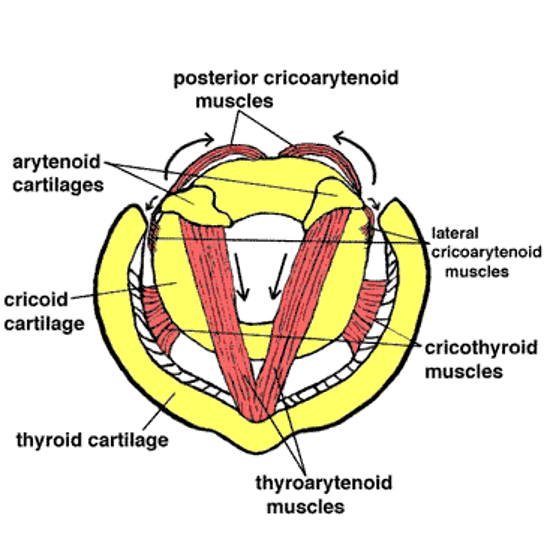
Figure 6: This is a view from above the larynx. In this view, muscles and cartilage are exposed. Arrows indicate the direction each muscle contracts.
Extrinsic Laryngeal Muscles
These are the muscles of the front of the neck and jaw that surround the larynx. The muscles of the front of the neck are also collectively referred to as the "strap" muscles.
We won't provide detail on all the extrinsic laryngeal muscles, but there are many to give the head and jaw their wide variety of movements. These muscles can provide stability for the larynx during phonation.
Though each of the strap muscles is responsible for a single specific movement, when there's tension in the neck, the muscles tend to contract as a unit (all at once). This tension can make it harder for the intrinsic laryngeal muscles to do their job. Extrinsic laryngeal muscle tension is a factor in many voice disorders (see Specific Voice Disorders).
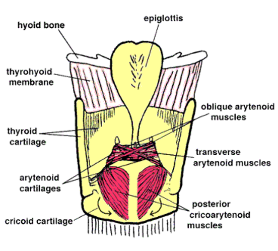
Figure 7: This is a view from behind the larynx. In this view, muscles and cartilage are exposed.
More Terms
- Phonation - Phonation is the word we use for making noise with the larynx, or producing vocal sound; phon- is a root word meaning sound.
- Dysphonia - Dysphonia is poor voice quality; dys- means bad, and phon- means sound. We often use the term dysphonia even if the sound is acceptable but the person has discomfort while phonating.
- Adduction - Adduction occurs when the vocal folds come together to close the glottis (think of adding the vocal folds together to remember this term).
- Abduction - Abduction is bringing the vocal folds apart to open the glottis for breathing
- Mucosa - Mucosa is the kind of tissue that lines the entire inside of the mouth, throat, etc. It is soft and wet, and should always be covered by a layer of secretions (saliva). The mucosal covering of the vocal folds is very special: it is made of several layers of collagen fibers. Each layer is arranged differently in order to give different kinds of strength yet flexibility for vibration. In most phonation, it is the mucosa that vibrates, not the entire vocal fold. See Figure 9.
For Your Information
You will often hear or read the term, “mucosa” in an ENT clinic. Mucosa is a generic term for the internal lining of our bodies, such as the insides of our cheeks and noses. It’s pink and it’s slimy. The vocal fold mucosa, however, is white. This is because there are many collagen and elastin fibers in the mucosa. This is good because if the vocal folds become irritated, red, and inflamed, they will repair themselves when the irritation stops. Mucus, on the other hand, is a term that refers to secretions produced in the upper airway. (You may call it phlegm) Those secretions are very important for the health of the mucosa. In a healthy larynx, the secretions are thin, and are swallowed without being noticed. (We swallow a quart of secretions every day!) In an irritated larynx, the secretions can become dry and thick, leading to the desire to cough or throat clear.
- Vocal Tract - This term refers to everything from the glottis to the lips. The vocal tract is the passage for the sound wave. Most people call it their throat! The vocal tract and the pharynx are essentially the same.
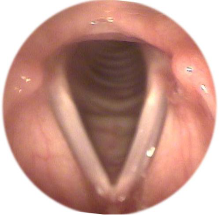
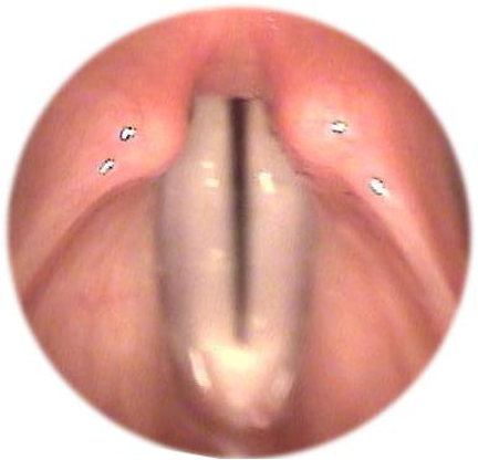
Figure 8A: A picture of abduction as we see it through an endoscope.
Figure 8B: A picture of adduction as we see it through an endoscope.
For Your Information
Here's something you probably never thought of…
Your larynx is called your voice box, but phonation (production of vocal sound) isn't the primary biological purpose of the larynx.
The most important thing your larynx does is . . . protect your lungs from food and water. The vocal folds form a valve that closes tightly to protect the airway. When you swallow, the glottis closes tightly—that is, the vocal folds adduct (come together) tightly. Also, the epiglottis folds over the glottis, and the larynx rises up while the esophagus opens to let the food/water enter. You've probably gotten water "down the wrong pipe" sometime in your life. That's because the water went down too fast for the glottis and epiglottis to protect the airway. When the water hit the vocal folds, they went into a cough reflex to expel the water and keep it out of the lungs. (Food or water in your lungs is a very bad thing.)
The second most important thing your larynx does is . . . trap air inside your lungs. This provides what we call "thoracic fixation," also known as the Valsalva maneuver. That is, trapping air inside the lungs provides air pressure, against which you push for excretion and childbirth, or stabilization for lifting.
We share these laryngeal attributes with air-breathing animals. The fact that our larynx can also make beautiful sounds is one of our very special human attributes.
Structure of the Vocal Fold
Vocal folds used to be called vocal cords (and are still often referred to that way) because it was thought that they vibrated much like strings on a violin. Also, from above, as seen from the laryngeal mirror, they do look like little pieces of cord. Now, we know more about them.
The figure below shows the cross section of a vocal fold.
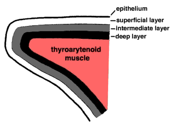
Figure 9: This drawing shows a cross section of a vocal fold. Deeper layers of mucosa are more stiff than shallower ones. At the core is a muscle with fibers stretching from front to back.
As you can see, it is a multilayered structure. The innermost layer is the thyroarytenoid (a.k.a. vocalis) muscle, which runs the entire length of the vocal fold, from the thyroid cartilage to the arytenoid cartilage. The thyroarytenoid muscle is the most dense portion of the vocal fold.
Surrounding the thyroarytenoid muscle is a sheath of mucosal tissue, that varies in stiffness from the stiffest portion surrounding the muscle, to the outermost layer, which is quite floppy.
The mucosal covering is divided into three sections, deep, intermediate, and superficial. All three layers are composed of collagen fibers, but the fibers are arranged differently in each layer. The fibers of the superficial layer are sparsely arranged, and are like thin, loose threads. The intermediate fibers are arranged along a different direction, and are more like a bundle of soft rubber bands. The fibers of the deep layer lie in yet a different direction, and they are arranged in dense, stiff bundles, like clumps of cotton thread. The overall looseness or stiffness of the mucosa depends on the state of contraction of the laryngeal muscles, but in general, the mucosa is similar in consistency to soft-set Jell-O.
The mucosa is covered by a thin layer of epithelial covering, similar to the loose skin on the back of your hand. This epithelium contains the mucosa but allows it to take on a variety of shapes.
The interesting thing about the multilayered structure is that the vocal fold can be stretched or contracted and made to vibrate at many different lengths. It can also adapt to different degrees of impact, and can withstand the forces and strain of extremely rapid vibration by rapidly changing its configuration.
Definitions
laryngeal [pronounced lah-RIN-jul or lair-in-JEE-al]: having to do with the larynx.
Abduction: The vocal folds abduct (come apart) in order to let air in and out of the lungs during breathing.
Adduction: The vocal folds may adduct (come together) to trap air in the lungs. They may also adduct to vibrate to produce vocal sound.
Interesting Facts
Do Women Have an Adam’s Apple? Both men and women have a thyroid notch, so both men and women have an Adam’s Apple. But men’s are more angular, and women’s are more rounded, and therefore less prominent.
—
Length of the vocal folds: The maximum length of the vocal folds is about 16 mm for an adult male and about 10 mm for an adult female.