Neuro-immuno Interface Lab
Expertise
Tissue repair and remodeling involving neuro-immuno interaction; Peripheral sensory disorder especially itch; Skin differentiation and proliferation control; Peripheral tissue circadian clock principle, mechanism, and control; Chronotherapy; 3D epithelia equivalent and epithelia-on-Chip.
Lab technique and tools
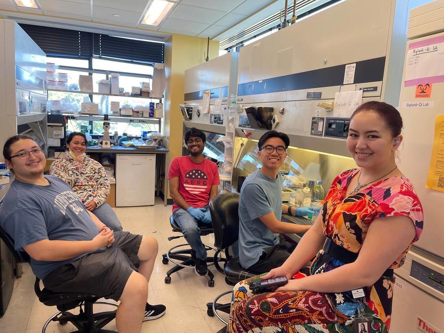
Dr. Bigliardi-Qi’s lab uses various molecular and cellular biology, pharmacology, and biomedical engineering including 3D printing techniques in both mechanistic studies and translational research projects. The human primary keratinocyte, stem cell-derived sensory neurons, immune and related cells are used for 2D, 3D, and co-culture models, epithelia-on-chip. RNA, DNA, and protein from human tissues such as epithelial, skin tissues, and PBMC are isolated and analyzed using RNAseq, qPCR, ChIP assay, western blot, ELISA, and EliSpot. Additionally, different microscopy and immuno- histochemistry are used for morphology and spatiotemporal biomarkers, and functional analysis. Bio-printing epithelial models and 3D printing Chips have been achieved together with the Earl Bakken medical device center. Novel in-vivo human studies have been conducted using photoacoustic and/or in-vivo Raman confocal microscopy.
Research Interest
The long-term goal of Dr. Bigliardi-Qi’s neuro-immuno interface lab is to understand the molecular mechanisms underlying epithelial tissue regeneration, differentiation and immune function, and skin keratinocyte involvement in skin sensory, tissue repair, and immunological disorder. The peripheral tissue clock is exposed to light and external environmental factors, making it a very important aspect of skin disorders. Her lab has been working on G-protein coupled receptors, their signaling pathways, circadian rhythm, and finally, photobiology and their epithelial applications.
Using opioid receptor knockout mice and human chronic pruritus trials, her lab has demonstrated that chronic itch is not histamine-mediated, but rather a neuronal and immune disorder. Regulation of epidermal m-opiate receptor (MOPr) improves itching, which is a major problem for aged skin with age-related peripheral neuropathy. Activation of the d-opiate receptor (DOPr) significantly improves wound healing in rodent chronic wound models. Her lab discovered various taste and olfactory receptors in skin epithelial cells and demonstrated that they act as important chemical sensors.
In addition, her lab developed both 2D co-cultures and award-winning 3D model systems of skin including the use of microfluidic systems (AAALAC award 2018). These systems were applied to analyze skin layers and structure for skin pharmacology using modern non-invasive methods (Raman spectroscopy, multispectral optoacoustic tomography, or MSOT). Clinical studies using these non-invasive methods led to the discovery of PPARγ‘s role regarding signaling in hair loss and evaluation of the microbiome in chronic, non-healing wounds. These activities led to several patents for novel therapies and screening devices. The 3D skin models her group developed included advanced melanoma and standardized wound models. This skin model can be expanded to other epithelial model cultures, e.g. oral, respiratory, or gut epithelium. These models could be used for toxicology and drug screening. Her lab has extensive experience working with multidisciplinary teams including engineers, clinicians, and basic research scientists to deliver novel translational research results such as drug targets, medical devices, products, and testing models. This expertise also is used to further develop novel treatments for specific epithelial disorders and other diseases to advance these therapies to clinical trials by partnering with the industry.
Since the COVID-19 pandemic, her lab has explored designing, developing, refurbishing, and adapting the EliSpot to the SARS-CoV2 conditions by testing different cytokines. The preliminary comparison indicated the cytokine pattern difference between asymptomatic and symptomatic. patient. This pilot study reveals that hyperactivation of PBMCs produces more cytokines after spike protein activation compared to a less strong activation.
Recently, her lab has discovered novel local photoentrainment mechanisms independent of the central clock. Furthermore, they proved that BMAL1/CLOCK acts as transcription factors to bind to promoters of important immune mediators. Finally, they confirmed the CR-inflammation signal pathway. This has a great impact on chronotherapy and great potential regarding improvement in the treatment of diseases with circadian aggravation.
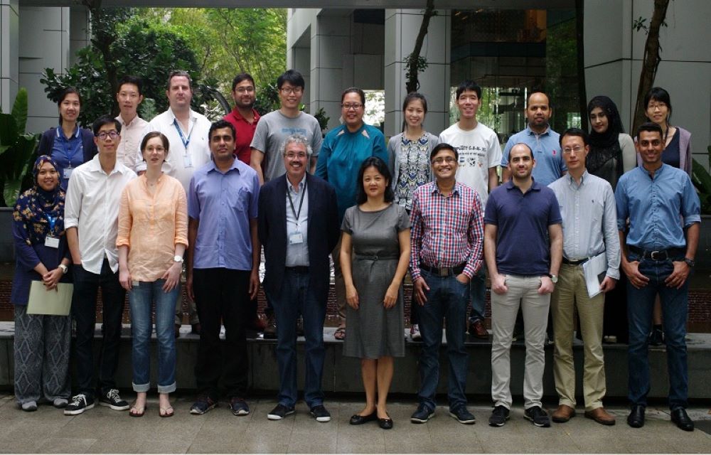
Previous lab: 2011-2017 CRUSAR (Clinical research unit for skin, allergy & regeneration)
