3D-printed Transparent Skull Provides a Window to the Brain
MINNEAPOLIS, MN- April 11, 2019- University of Minnesota researchers have developed a 3D-printed transparent skull implant that could provide new insight into how complex brain networks function and to understand abnormal conditions such as concussions, Alzheimer’s and Parkinson’s disease.
In new research is published in Nature Communications, Timothy J. Ebner, MD, PhD, a co-author of the study and a University of Minnesota Professor and Head of the Department of Neuroscience in the Medical School, discusses how the study, currently done only in mice, provides an opportunity to watch the activity of the entire brain surface in real time.
“These are studies that are extremely important in our understanding of how the brain works normally and in disease processes, with the long term goal of improving treatments for people who experience brain injuries or diseases,” said Ebner.
This sort of understanding has never been reachable before, because scientists lacked the tools to allow simultaneous monitoring and perturbing neural activity from multiple cortical regions. This device, called See-Shell, allows researchers to see global changes for the first time at an unprecedented time resolution.
“What we are trying to do is to see if we can visualize and interact with large parts of the mouse brain surface, called the cortex, over long periods of time. This will give us new information about how the human brain works,” said Suhasa Kodandaramaiah, PhD, the senior author of the study and University of Minnesota Benjamin Mayhugh Assistant Professor of Mechanical Engineering in the College of Science and Engineering. “This technology allows us to see most of the cortex in action with unprecedented control and precision while stimulating certain parts of the brain.”
One of their first studies using the See-Shell device examines how mild concussions in one part of the brain affect other parts of the brain as it reorganizes structurally and functionally. Kodandaramaiah said that mouse brains are very similar in many respects to human brains, and this device opens the door for similar research on mice looking at degenerative brain diseases that affect humans such as Alzheimer’s or Parkinson’s disease.
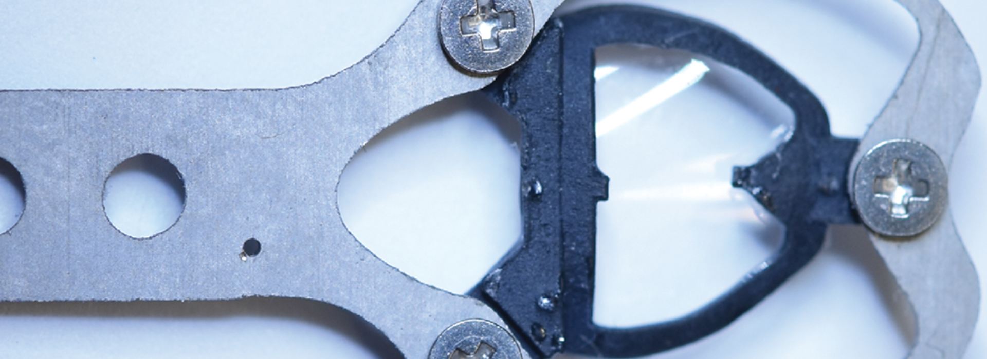
To make the See-Shell, researchers digitally scanned the surface of the mouse skull and then used the digital scans to create an artificial transparent skull that has the same contours as the original skull. During a precise surgery, the top of the mouse skull is replaced with the 3D-printed transparent skull device. The device allows researchers to record brain activity simultaneously while imaging the entire brain in real time.
Another advantage to using this device is that researchers were able to study the same brain over several months. Studies in mice over several months allow researchers to study brain aging in a way that would take decades to study in humans.
The research team was led by fourth-year mechanical engineering PhD student Leila Ghanbari. The research team included several post-doctoral associates, graduate students and undergraduate students including Russell E. Carter (neuroscience), Matthew L. Rynes (biomedical engineering), Judith Dominguez (mechanical engineering), Gang Chen (neuroscience), Anant Naik (biomedical engineering), Jia Hu (biomedical engineering), Lenora Haltom (mechanical engineering), Nahom Mossazghi (neuroscience), Madelyn M. Gray (neuroscience) and Sarah L. West (neuroscience). The team also included partners at the University of Wisconsin including researcher Kevin W. Eliceiri and graduate student Md Abdul Kader Sagar.
The research was funded primarily by the National Institutes of Health (NIH) with additional support from Minnesota’s Discovery, Research, and InnoVation Economy (MnDRIVE) funding from the State of Minnesota. Several undergraduate students involved in the research were supported by the University of Minnesota Undergraduate Research Opportunities Program (UROP). The imaging research was made possible by the cutting-edge imaging infrastructure at the University Imaging Center.
Read the full research paper, entitled “Cortex-wide neural interfacing via transparent polymer skulls,” here.